ARG63748
anti-ACHE antibody
anti-ACHE antibody for Flow cytometry,ICC/IF,Western blot and Human
Neuroscience antibody
Overview
| Product Description | Goat Polyclonal antibody recognizes ACHE |
|---|---|
| Tested Reactivity | Hu |
| Predict Reactivity | Ms, Rat |
| Tested Application | FACS, ICC/IF, WB |
| Specificity | This antibody is expected to recognise isoform NP_000656 only (the ubiquitously expressed, hydrophillic form). |
| Host | Goat |
| Clonality | Polyclonal |
| Isotype | IgG |
| Target Name | ACHE |
| Antigen Species | Human |
| Immunogen | QFDHYSKQDRCSDL |
| Conjugation | Un-conjugated |
| Alternate Names | ARACHE; Acetylcholinesterase; ACEE; EC 3.1.1.7; AChE; N-ACHE; YT |
Application Instructions
| Application Suggestion |
|
||||||||
|---|---|---|---|---|---|---|---|---|---|
| Application Note | WB: Recommend incubate at RT for 1h. * The dilutions indicate recommended starting dilutions and the optimal dilutions or concentrations should be determined by the scientist. |
Properties
| Form | Liquid |
|---|---|
| Purification | Purified from goat serum by antigen affinity chromatography. |
| Buffer | Tris saline (pH 7.3), 0.02% Sodium azide and 0.5% BSA. |
| Preservative | 0.02% Sodium azide |
| Stabilizer | 0.5% BSA |
| Concentration | 0.5 mg/ml |
| Storage Instruction | For continuous use, store undiluted antibody at 2-8°C for up to a week. For long-term storage, aliquot and store at -20°C or below. Storage in frost free freezers is not recommended. Avoid repeated freeze/thaw cycles. Suggest spin the vial prior to opening. The antibody solution should be gently mixed before use. |
| Note | For laboratory research only, not for drug, diagnostic or other use. |
Bioinformation
| Database Links | |
|---|---|
| Background | Acetylcholinesterase hydrolyzes the neurotransmitter, acetylcholine at neuromuscular junctions and brain cholinergic synapses, and thus terminates signal transmission. It is also found on the red blood cell membranes, where it constitutes the Yt blood group antigen. Acetylcholinesterase exists in multiple molecular forms which possess similar catalytic properties, but differ in their oligomeric assembly and mode of cell attachment to the cell surface. It is encoded by the single ACHE gene, and the structural diversity in the gene products arises from alternative mRNA splicing, and post-translational associations of catalytic and structural subunits. The major form of acetylcholinesterase found in brain, muscle and other tissues is the hydrophilic species, which forms disulfide-linked oligomers with collagenous, or lipid-containing structural subunits. The other, alternatively spliced form, expressed primarily in the erythroid tissues, differs at the C-terminal end, and contains a cleavable hydrophobic peptide with a GPI-anchor site. It associates with the membranes through the phosphoinositide (PI) moieties added post-translationally. [provided by RefSeq, Jul 2008] |
| Research Area | Neuroscience antibody |
| Calculated MW | 68 kDa |
Images (3) Click the Picture to Zoom In
-
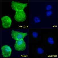
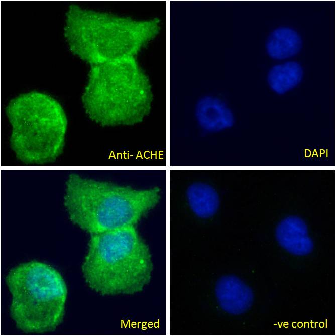
ARG63748 anti-ACHE antibody ICC/IF image
Immunofluorescence: Paraformaldehyde fixed U2OS cells permeabilized with 0.15% Triton. Cells were stained with ARG63748 anti-ACHE antibody (green) at 10 µg/ml dilution for 1 hour. DAPI (blue) for nuclear staining. Negative control: Unimmunized goat IgG (green) at 10 µg/ml dilution.
-

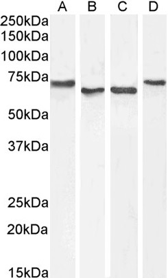
ARG63748 anti-ACHE antibody WB image
Western blot: 35 µg of HepG2 (A), Daudi (B), HeLa (C) and Jurkat (D) cell lysates (in RIPA buffer) stained with ARG63748 anti-ACHE antibody at 1 µg/ml (A, B) and 0.3 µg/ml (C, D) dilutions and incubated at RT for 1 hour.
-
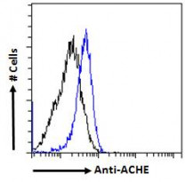
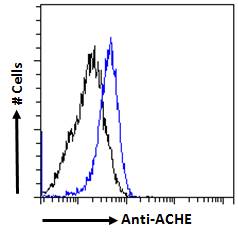
ARG63748 anti-ACHE antibody FACS image
Flow Cytometry: Paraformaldehyde-fixed HeLa cells permeabilized with 0.5% Triton. Cells were stained with ARG63748 anti-ACHE antibody (blue line) at 10 µg/ml dilution for 1 hour, followed by incubation with Alexa FluorR 488 labelled secondary antibody. IgG control: Unimmunized goat IgG (black line).