ARG55283
anti-ACHE antibody
anti-ACHE antibody for ICC/IF,Western blot and Human,Mouse,Rat
Neuroscience antibody
Overview
| Product Description | Rabbit Polyclonal antibody recognizes ACHE |
|---|---|
| Tested Reactivity | Hu, Ms, Rat |
| Tested Application | ICC/IF, WB |
| Host | Rabbit |
| Clonality | Polyclonal |
| Isotype | IgG |
| Target Name | ACHE |
| Antigen Species | Human |
| Immunogen | Recombinant protein of Human ACHE |
| Conjugation | Un-conjugated |
| Alternate Names | ARACHE; Acetylcholinesterase; ACEE; EC 3.1.1.7; AChE; N-ACHE; YT |
Application Instructions
| Application Suggestion |
|
||||||
|---|---|---|---|---|---|---|---|
| Application Note | * The dilutions indicate recommended starting dilutions and the optimal dilutions or concentrations should be determined by the scientist. | ||||||
| Positive Control | Rat spinal cord, Mouse brain and SH-SY5Y | ||||||
| Observed Size | 75 kDa |
Properties
| Form | Liquid |
|---|---|
| Purification | Affinity purification with immunogen. |
| Buffer | PBS (pH 7.3), 0.02% Sodium azide and 50% Glycerol |
| Preservative | 0.02% Sodium azide |
| Stabilizer | 50% Glycerol |
| Storage Instruction | For continuous use, store undiluted antibody at 2-8°C for up to a week. For long-term storage, aliquot and store at -20°C. Storage in frost free freezers is not recommended. Avoid repeated freeze/thaw cycles. Suggest spin the vial prior to opening. The antibody solution should be gently mixed before use. |
| Note | For laboratory research only, not for drug, diagnostic or other use. |
Bioinformation
| Database Links | |
|---|---|
| Gene Symbol | ACHE |
| Gene Full Name | acetylcholinesterase (Yt blood group) |
| Background | Acetylcholinesterase hydrolyzes the neurotransmitter, acetylcholine at neuromuscular junctions and brain cholinergic synapses, and thus terminates signal transmission. It is also found on the red blood cell membranes, where it constitutes the Yt blood group antigen. Acetylcholinesterase exists in multiple molecular forms which possess similar catalytic properties, but differ in their oligomeric assembly and mode of cell attachment to the cell surface. It is encoded by the single ACHE gene, and the structural diversity in the gene products arises from alternative mRNA splicing, and post-translational associations of catalytic and structural subunits. The major form of acetylcholinesterase found in brain, muscle and other tissues is the hydrophilic species, which forms disulfide-linked oligomers with collagenous, or lipid-containing structural subunits. The other, alternatively spliced form, expressed primarily in the erythroid tissues, differs at the C-terminal end, and contains a cleavable hydrophobic peptide with a GPI-anchor site. It associates with the membranes through the phosphoinositide (PI) moieties added post-translationally. [provided by RefSeq, Jul 2008] |
| Function | Terminates signal transduction at the neuromuscular junction by rapid hydrolysis of the acetylcholine released into the synaptic cleft. Role in neuronal apoptosis. [UniProt] |
| Research Area | Neuroscience antibody |
| Calculated MW | 68 kDa |
Images (2) Click the Picture to Zoom In
-
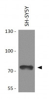
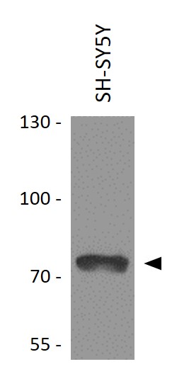
ARG55283 anti-ACHE antibody WB image
Western blot: 25 µg of SH-SY5Y cell lysate stained with ARG55283 anti-ACHE antibody at 1:1000 dilution.
-
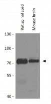
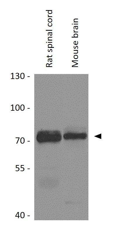
ARG55283 anti-ACHE antibody WB image
Western blot: 25 µg of Rat spinal cord and Mouse brain lysates stained with ARG55283 anti-ACHE antibody at 1:1000 dilution.