ARG57040
anti-ACAT2 antibody [55D5]
anti-ACAT2 antibody [55D5] for Flow cytometry,ICC/IF,Western blot and Human
Overview
| Product Description | Mouse Monoclonal antibody [55D5] recognizes ACAT2 |
|---|---|
| Tested Reactivity | Hu |
| Tested Application | FACS, ICC/IF, WB |
| Host | Mouse |
| Clonality | Monoclonal |
| Clone | 55D5 |
| Isotype | IgG2b, kappa |
| Target Name | ACAT2 |
| Antigen Species | Human |
| Immunogen | Recombinant fragment around aa. 1-397 of Human ACAT2. |
| Conjugation | Un-conjugated |
| Alternate Names | Acetyl-CoA transferase-like protein; EC 2.3.1.9; Acetyl-CoA acetyltransferase, cytosolic; Cytosolic acetoacetyl-CoA thiolase |
Application Instructions
| Application Suggestion |
|
||||||||
|---|---|---|---|---|---|---|---|---|---|
| Application Note | * The dilutions indicate recommended starting dilutions and the optimal dilutions or concentrations should be determined by the scientist. |
Properties
| Form | Liquid |
|---|---|
| Purification | Purification with Protein A. |
| Buffer | PBS (pH 7.4), 0.02% Sodium azide and 10% Glycerol. |
| Preservative | 0.02% Sodium azide |
| Stabilizer | 10% Glycerol |
| Concentration | 1 mg/ml |
| Storage Instruction | For continuous use, store undiluted antibody at 2-8°C for up to a week. For long-term storage, aliquot and store at -20°C. Storage in frost free freezers is not recommended. Avoid repeated freeze/thaw cycles. Suggest spin the vial prior to opening. The antibody solution should be gently mixed before use. |
| Note | For laboratory research only, not for drug, diagnostic or other use. |
Bioinformation
| Database Links |
Swiss-port # Q9BWD1 Human Acetyl-CoA acetyltransferase, cytosolic |
|---|---|
| Gene Symbol | ACAT2 |
| Gene Full Name | acetyl-CoA acetyltransferase 2 |
| Background | The product of this gene is an enzyme involved in lipid metabolism, and it encodes cytosolic acetoacetyl-CoA thiolase. This gene shows complementary overlapping with the 3-prime region of the TCP1 gene in both mouse and human. These genes are encoded on opposite strands of DNA, as well as in opposite transcriptional orientation. Alternatively spliced transcript variants encoding different isoforms have been found for this gene. [provided by RefSeq, Dec 2014] |
| Calculated MW | 41 kDa |
Images (5) Click the Picture to Zoom In
-
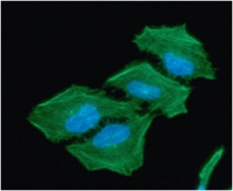
ARG57040 anti-ACAT2 antibody [55D5] ICC/IF image
Immunoflorescense: HeLa cell line stained with ARG57040 anti-ACAT2 antibody [55D5] at 1:100 (Green).
DAPI (Blue) for nucleus staining.
-
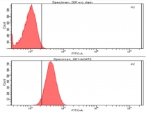
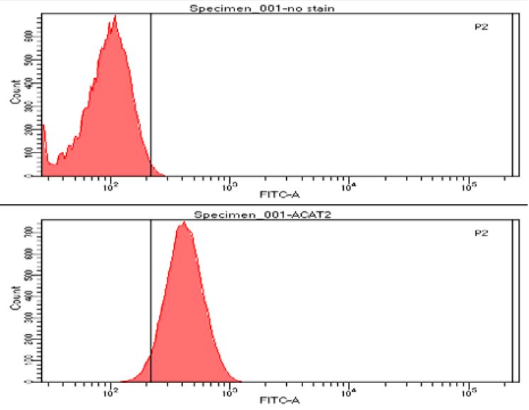
ARG57040 anti-ACAT2 antibody [55D5] FACS image
Flow Cytometry: PC3 cell line stained with ARG57040 anti-ACAT2 antibody [55D5] at 2-5 µg for 1x10^6 cells. Secondary antibody: Goat antiMouse IgG Alexa fluor 488 conjugate.
-
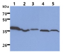
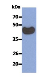
ARG57040 anti-ACAT2 antibody [55D5] WB image
Western blot: 40 µg of 1) HepG2 cell lysate, 2) Ramos cell lysate, 3) MCF-7 cell lysate, 4) HL-60 cell lysate, 5) SW480 cell lysate stained with ARG57040 anti-ACAT2 antibody [55D5] at 1:1000.
-
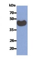
ARG57040 anti-ACAT2 antibody [55D5] WB image
Western blot: 50 ng of Recombinant Human ACAT2 stained with ARG57040 anti-ACAT2 antibody [55D5] at 1:1000.
-

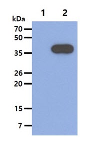
ARG57040 anti-ACAT2 antibody [55D5] WB image
Western blot: 5 µg of 1) 293T cell lysate, 2) ACAT2 Transfected 293T cell lysate stained with ARG57040 anti-ACAT2 antibody [55D5] at 1:1000.