ARG66308
anti-ACAA2 antibody
anti-ACAA2 antibody for IHC-Formalin-fixed paraffin-embedded sections,Western blot and Human,Mouse
Overview
| Product Description | Rabbit Polyclonal antibody recognizes ACAA2 |
|---|---|
| Tested Reactivity | Hu, Ms |
| Tested Application | IHC-P, WB |
| Host | Rabbit |
| Clonality | Polyclonal |
| Isotype | IgG |
| Target Name | ACAA2 |
| Antigen Species | Human |
| Immunogen | Fusion protein of Human ACAA2. |
| Conjugation | Un-conjugated |
| Alternate Names | Acetyl-CoA acyltransferase; Beta-ketothiolase; T1; EC 2.3.1.16; DSAEC; 3-ketoacyl-CoA thiolase, mitochondrial; Mitochondrial 3-oxoacyl-CoA thiolase |
Application Instructions
| Application Suggestion |
|
||||||
|---|---|---|---|---|---|---|---|
| Application Note | * The dilutions indicate recommended starting dilutions and the optimal dilutions or concentrations should be determined by the scientist. | ||||||
| Positive Control | WB: NIH/3T3 and A172 cells, Human liver cancer tissue. IHC-P: Human colon cancer and Human thyroid cancer. |
Properties
| Form | Liquid |
|---|---|
| Purification | Affinity purification with immunogen. |
| Buffer | PBS (pH 7.4), 0.05% Sodium azide and 40% Glycerol. |
| Preservative | 0.05% Sodium azide |
| Stabilizer | 40% Glycerol |
| Concentration | 1.5 mg/ml |
| Storage Instruction | For continuous use, store undiluted antibody at 2-8°C for up to a week. For long-term storage, aliquot and store at -20°C. Storage in frost free freezers is not recommended. Avoid repeated freeze/thaw cycles. Suggest spin the vial prior to opening. The antibody solution should be gently mixed before use. |
| Note | For laboratory research only, not for drug, diagnostic or other use. |
Bioinformation
| Database Links |
Swiss-port # P42765 Human 3-ketoacyl-CoA thiolase, mitochondrial Swiss-port # Q8BWT1 Mouse 3-ketoacyl-CoA thiolase, mitochondrial |
|---|---|
| Gene Symbol | ACAA2 |
| Gene Full Name | acetyl-CoA acyltransferase 2 |
| Background | The encoded protein catalyzes the last step of the mitochondrial fatty acid beta-oxidation spiral. Unlike most mitochondrial matrix proteins, it contains a non-cleavable amino-terminal targeting signal. [provided by RefSeq, Jul 2008] |
| Function | Abolishes BNIP3-mediated apoptosis and mitochondrial damage. [UniProt] |
| Calculated MW | 42 kDa |
Images (3) Click the Picture to Zoom In
-
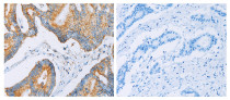
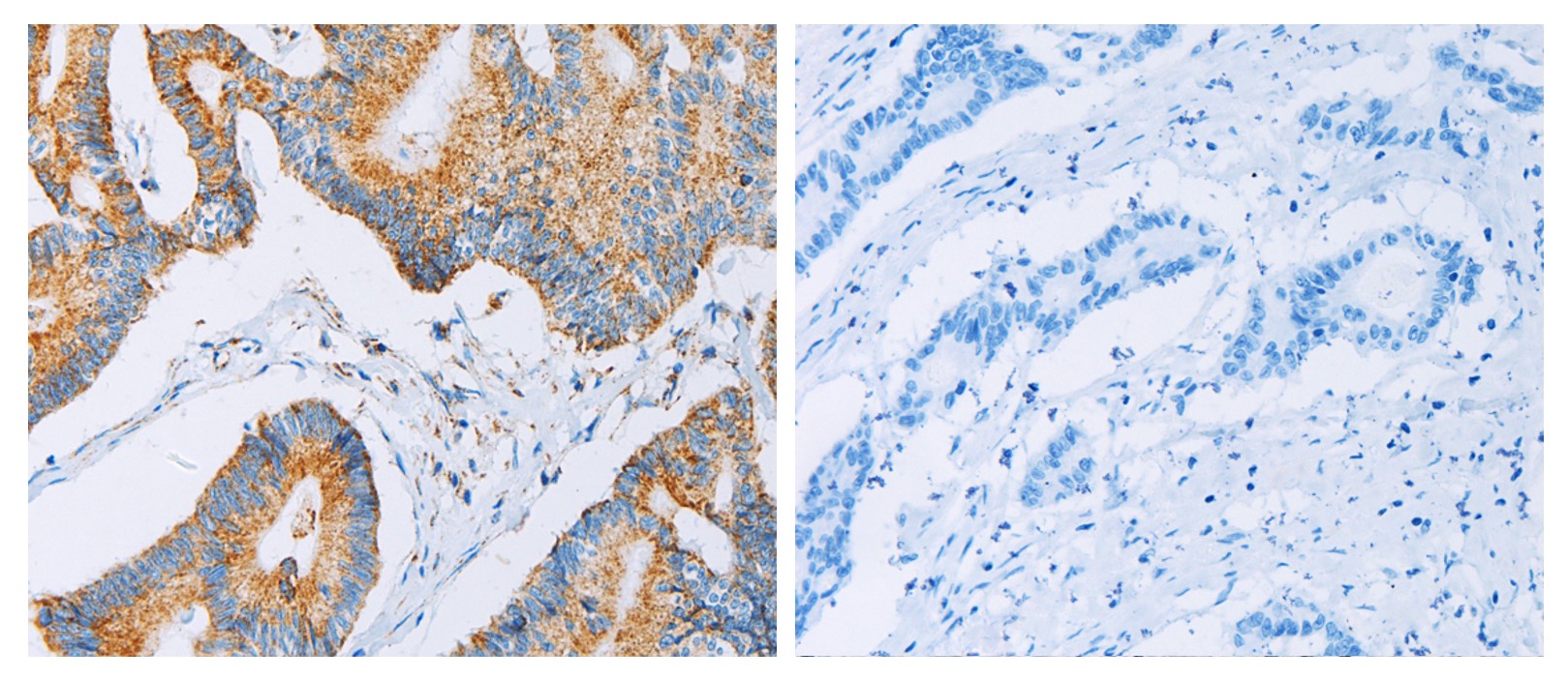
ARG66308 anti-ACAA2 antibody IHC-P image
Immunohistochemistry: Paraffin-embedded Human colon cancer tissue stained with ARG66308 anti-ACAA2 antibody (left) at 1:30 dilution, or the same antibody pre-incubated with antigen (right). (Original magnification: X200) .
-

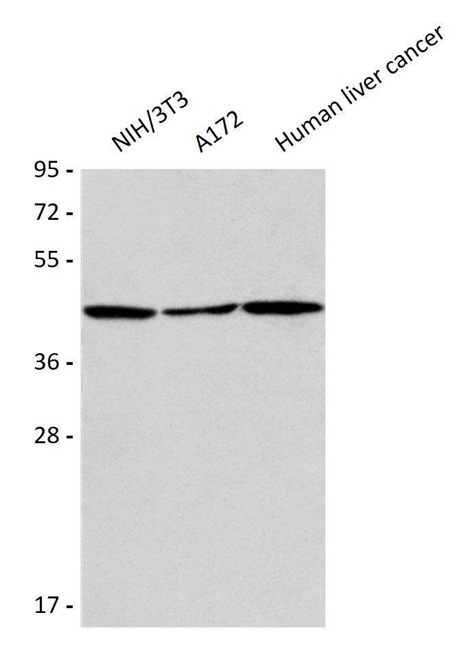
ARG66308 anti-ACAA2 antibody WB image
Western blot: 40 µg of NIH/3T3, A172 and Human liver cancer lysates stained with ARG66308 anti-ACAA2 antibody at 1:375 dilution.
-
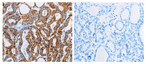
ARG66308 anti-ACAA2 antibody IHC-P image
Immunohistochemistry: Paraffin-embedded Human thyroid cancer tissue stained with ARG66308 anti-ACAA2 antibody (left) at 1:30 dilution, or the same antibody pre-incubated with antigen (right). (Original magnification: X200) .