ARG66172
anti-ABCB5 antibody
anti-ABCB5 antibody for IHC-Formalin-fixed paraffin-embedded sections,Western blot and Human
Overview
| Product Description | Mouse Monoclonal antibody recognizes ABCB5 |
|---|---|
| Tested Reactivity | Hu |
| Tested Application | IHC-P, WB |
| Specificity | The antibody detects endogenous ABCB5 proteins. |
| Host | Mouse |
| Clonality | Monoclonal |
| Target Name | ABCB5 |
| Antigen Species | Human |
| Immunogen | Synthetic peptide of Human ABCB5. |
| Conjugation | Un-conjugated |
| Alternate Names | ATP-binding cassette sub-family B member 5; ABCB5 P-gp; P-glycoprotein ABCB5; ABCB5beta; EST422562; ABCB5alpha |
Application Instructions
| Application Suggestion |
|
||||||
|---|---|---|---|---|---|---|---|
| Application Note | IHC-P: Antigen Retrieval: Boil tissue section in Sodium citrate buffer (pH 6.0) for 20 min. * The dilutions indicate recommended starting dilutions and the optimal dilutions or concentrations should be determined by the scientist. |
Properties
| Form | Liquid |
|---|---|
| Purification | Affinity purification with immunogen. |
| Buffer | PBS (pH 7.4), 0.02% Sodium azide and 50% Glycerol. |
| Preservative | 0.02% Sodium azide |
| Stabilizer | 50% Glycerol |
| Concentration | 1 mg/ml |
| Storage Instruction | For continuous use, store undiluted antibody at 2-8°C for up to a week. For long-term storage, aliquot and store at -20°C. Storage in frost free freezers is not recommended. Avoid repeated freeze/thaw cycles. Suggest spin the vial prior to opening. The antibody solution should be gently mixed before use. |
| Note | For laboratory research only, not for drug, diagnostic or other use. |
Bioinformation
| Database Links |
Swiss-port # Q2M3G0 Human ATP-binding cassette sub-family B member 5 |
|---|---|
| Gene Symbol | ABCB5 |
| Gene Full Name | ATP-binding cassette, sub-family B (MDR/TAP), member 5 |
| Background | ABCB5 belongs to the ATP-binding cassette (ABC) transporter superfamily of integral membrane proteins. These proteins participate in ATP-dependent transmembrane transport of structurally diverse molecules ranging from small ions, sugars, and peptides to more complex organic molecules (Chen et al., 2005 [PubMed 15760339]).[supplied by OMIM, Mar 2008] |
| Function | Drug efflux transporter present in a number of stem cells that acts as a regulator of cellular differentiation. Able to mediate efflux from cells of the rhodamine dye and of the therapeutic drug doxorubicin. Specifically present in limbal stem cells, where it plays a key role in corneal development and repair. [UniProt] |
| Calculated MW | 139 kDa |
Images (13) Click the Picture to Zoom In
-
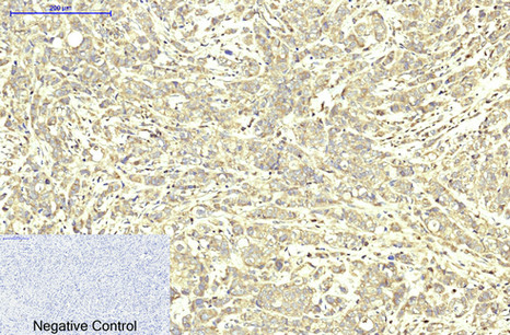
ARG66172 anti-ABCB5 antibody IHC-P image
Immunohistochemistry: Paraffin-embedded Human breast cancer tissue stained with ARG66172 anti-ABCB5 antibody at 1:200 dilution (4°C, overnight). Antigen Retrieval: Boil tissue section in Sodium citrate buffer (pH 6.0) for 20 min.
Negative control was used by secondary antibody only.
-
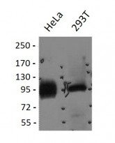
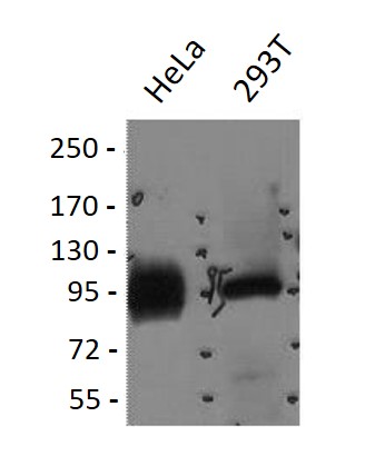
ARG66172 anti-ABCB5 antibody WB image
Western blot: 1) HeLa, and 2) 293T cell lysates stained with ARG66172 anti-ABCB5 antibody at 1:2000 dilution.
-
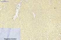
ARG66172 anti-ABCB5 antibody IHC-P image
Immunohistochemistry: Paraffin-embedded Human liver tissue stained with ARG66172 anti-ABCB5 antibody at 1:200 dilution (4°C, overnight). Antigen Retrieval: Boil tissue section in Sodium citrate buffer (pH 6.0) for 20 min.
Negative control was used by secondary antibody only.
-
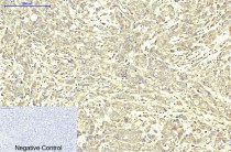
ARG66172 anti-ABCB5 antibody IHC-P image
Immunohistochemistry: Paraffin-embedded Human liver cancer tissue stained with ARG66172 anti-ABCB5 antibody at 1:200 dilution (4°C, overnight). Antigen Retrieval: Boil tissue section in Sodium citrate buffer (pH 6.0) for 20 min.
Negative control was used by secondary antibody only.
-
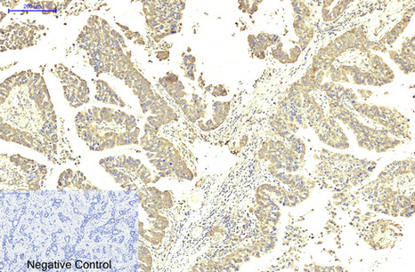
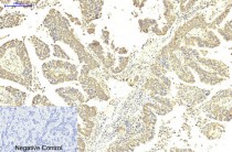
ARG66172 anti-ABCB5 antibody IHC-P image
Immunohistochemistry: Paraffin-embedded Human lung cancer tissue stained with ARG66172 anti-ABCB5 antibody at 1:200 dilution (4°C, overnight). Antigen Retrieval: Boil tissue section in Sodium citrate buffer (pH 6.0) for 20 min.
Negative control was used by secondary antibody only.
-
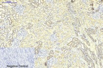
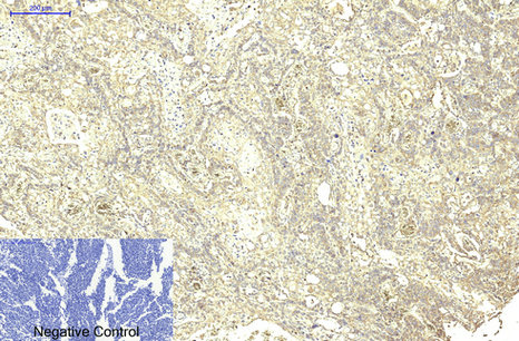
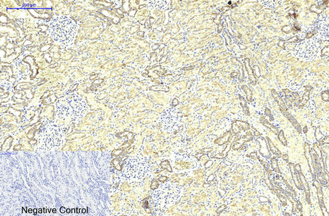
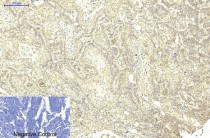
ARG66172 anti-ABCB5 antibody IHC-P image
Immunohistochemistry: Paraffin-embedded Human kidney tissue stained with ARG66172 anti-ABCB5 antibody at 1:200 dilution (4°C, overnight). Antigen Retrieval: Boil tissue section in Sodium citrate buffer (pH 6.0) for 20 min.
Negative control was used by secondary antibody only.
-

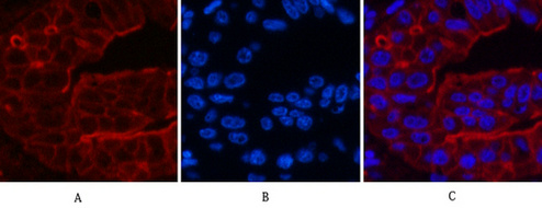
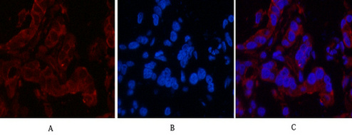
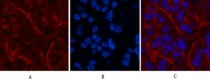
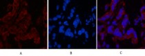
ARG66172 anti-ABCB5 antibody IHC image
Immunohistochemistry: Human liver cancer tissue stained with ARG66172 anti-ABCB5 antibody (red) at 1:200 dilution (4°C, overnight).
Picture A: Target. Picture B: DAPI. Picture C: merge of A+B.
-
ARG66172 anti-ABCB5 antibody IHC image
Immunohistochemistry: Human liver cancer tissue stained with ARG66172 anti-ABCB5 antibody (red) at 1:200 dilution (4°C, overnight).
Picture A: Target. Picture B: DAPI. Picture C: merge of A+B.
-
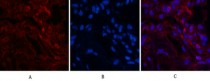
ARG66172 anti-ABCB5 antibody IHC image
Immunohistochemistry: Human liver cancer tissue stained with ARG66172 anti-ABCB5 antibody (red) at 1:200 dilution (4°C, overnight).
Picture A: Target. Picture B: DAPI. Picture C: merge of A+B.
-
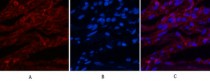
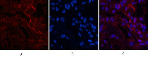
ARG66172 anti-ABCB5 antibody IHC image
Immunohistochemistry: Human lung tissue stained with ARG66172 anti-ABCB5 antibody (red) at 1:200 dilution (4°C, overnight).
Picture A: Target. Picture B: DAPI. Picture C: merge of A+B.
-
ARG66172 anti-ABCB5 antibody IHC image
Immunohistochemistry: Human lung tissue stained with ARG66172 anti-ABCB5 antibody (red) at 1:200 dilution (4°C, overnight).
Picture A: Target. Picture B: DAPI. Picture C: merge of A+B.
-
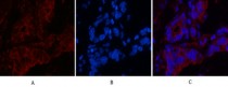
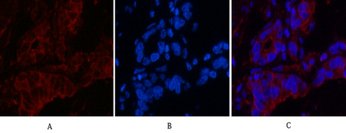
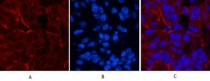
ARG66172 anti-ABCB5 antibody IHC image
Immunohistochemistry: Human stomach cancer tissue stained with ARG66172 anti-ABCB5 antibody (red) at 1:200 dilution (4°C, overnight).
Picture A: Target. Picture B: DAPI. Picture C: merge of A+B.
-
ARG66172 anti-ABCB5 antibody IHC image
Immunohistochemistry: Human stomach cancer tissue stained with ARG66172 anti-ABCB5 antibody (red) at 1:200 dilution (4°C, overnight).
Picture A: Target. Picture B: DAPI. Picture C: merge of A+B.