ARG57840
anti-ABAT antibody
anti-ABAT antibody for ICC/IF,Western blot and Human,Mouse,Rat
Overview
| Product Description | Rabbit Polyclonal antibody recognizes ABAT |
|---|---|
| Tested Reactivity | Hu, Ms, Rat |
| Tested Application | ICC/IF, WB |
| Host | Rabbit |
| Clonality | Polyclonal |
| Isotype | IgG |
| Target Name | ABAT |
| Antigen Species | Human |
| Immunogen | Recombinant protein of Human ABAT. |
| Conjugation | Un-conjugated |
| Alternate Names | GABAT; GABA-T; EC 2.6.1.22; GABA aminotransferase; GABA-AT; GABA transaminase; EC 2.6.1.19; NPD009; S; 4-aminobutyrate aminotransferase, mitochondrial; Gamma-amino-N-butyrate transaminase; L-AIBAT |
Application Instructions
| Application Suggestion |
|
||||||
|---|---|---|---|---|---|---|---|
| Application Note | * The dilutions indicate recommended starting dilutions and the optimal dilutions or concentrations should be determined by the scientist. | ||||||
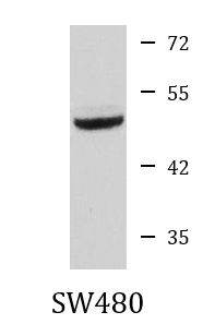
| Positive Control | SW480 |
Properties
| Form | Liquid |
|---|---|
| Purification | Affinity purified. |
| Buffer | PBS (pH 7.3), 0.02% Sodium azide and 50% Glycerol. |
| Preservative | 0.02% Sodium azide |
| Stabilizer | 50% Glycerol |
| Storage Instruction | For continuous use, store undiluted antibody at 2-8°C for up to a week. For long-term storage, aliquot and store at -20°C. Storage in frost free freezers is not recommended. Avoid repeated freeze/thaw cycles. Suggest spin the vial prior to opening. The antibody solution should be gently mixed before use. |
| Note | For laboratory research only, not for drug, diagnostic or other use. |
Bioinformation
| Database Links | |
|---|---|
| Gene Symbol | ABAT |
| Gene Full Name | 4-aminobutyrate aminotransferase |
| Background | 4-aminobutyrate aminotransferase (ABAT) is responsible for catabolism of gamma-aminobutyric acid (GABA), an important, mostly inhibitory neurotransmitter in the central nervous system, into succinic semialdehyde. The active enzyme is a homodimer of 50-kD subunits complexed to pyridoxal-5-phosphate. The protein sequence is over 95% similar to the pig protein. GABA is estimated to be present in nearly one-third of human synapses. ABAT in liver and brain is controlled by 2 codominant alleles with a frequency in a Caucasian population of 0.56 and 0.44. The ABAT deficiency phenotype includes psychomotor retardation, hypotonia, hyperreflexia, lethargy, refractory seizures, and EEG abnormalities. Multiple alternatively spliced transcript variants encoding the same protein isoform have been found for this gene. [provided by RefSeq, Jul 2008] |
| Function | Catalyzes the conversion of gamma-aminobutyrate and L-beta-aminoisobutyrate to succinate semialdehyde and methylmalonate semialdehyde, respectively. Can also convert delta-aminovalerate and beta-alanine. [UniProt] |
| Cellular Localization | Mitochondrion matrix. [UniProt] |
| Calculated MW | 56 kDa |
Images (2) Click the Picture to Zoom In
-
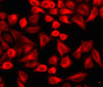
ARG57840 anti-ABAT antibody ICC/IF image
Immunofluorescence: A549 cells stained with ARG57840 anti-ABAT antibody.
-

ARG57840 anti-ABAT antibody WB image
Western blot: 25 µg of SW480 cell lysate stained with ARG57840 anti-ABAT antibody at 1:1000 dilution.