ARG30155
Phospho Smad1 for Activation / Inhibition site Antibody Panel
Cancer antibody; Developmental Biology antibody; Gene Regulation antibody; Immune System antibody; Metabolism antibody; Signaling Transduction antibody
Component
| Cat No | Component Name | Host clonality | Reactivity | Application | Package |
|---|---|---|---|---|---|
| ARG51284 | anti-Smad 1 antibody | Rabbit pAb | Hu, Ms, Rat | ICC/IF, WB | 50 μl |
| ARG51848 | anti-Smad 1 phospho (Ser206) antibody | Rabbit pAb | Hu, Ms, Rat | WB | 50 μl |
| ARG51794 | anti-Smad 1 phospho (Ser465) antibody | Rabbit pAb | Hu, Ms, Rat | WB | 50 μl |
| ARG65351 | Goat anti-Rabbit IgG antibody (HRP) | Goat pAb | Rb | ELISA, IHC-P, WB | 50 μl |
Overview
| Product Description | Smad transcription factors convey downstream messages from the most versatile cytokine signaling pathways in the multicellular life forms, the transforming growth factor-β (TGFβ) pathway. Recent works has shed light into the processes and functions of SMAD activation and deactivation, nucleocytoplasmic dynamics, and assembly of transcriptional complexes. Smad proteins consist of two globular domains coupled by a linker region. Smad1 can be phosphorylated at the linker region (pSer206) where recruitment of Smurf takes place and results in the degradation of Smad1. On the other hands, phosphorylation of Smad1 at pSer465 translocate Smad1/Smad4 complex to nucleus and transcriptionally activate target genes. Whiteman et al. (1993) Genes Dev 12:2445-62 Massagué J et al. (2005) Genes Dev 19: 2783-2810 Sapkota et al. (2007) Mol Cell 25: 441-454 |
|---|---|
| Target Name | Smad1 for Activation / Inhibition site |
| Alternate Names | Phospho Smad1 for Activation / Inhibition site antibody; Smad 1 antibody; Smad 1 phospho (Ser465) antibody; Smad 1 phospho (Ser206) antibody |
Properties
| Storage Instruction | For continuous use, store undiluted antibody at 2-8°C for up to a week. For long-term storage, aliquot and store at -20°C or below. Storage in frost free freezers is not recommended. Avoid repeated freeze/thaw cycles. Suggest spin the vial prior to opening. The antibody solution should be gently mixed before use. |
|---|---|
| Note | For laboratory research only, not for drug, diagnostic or other use. |
Bioinformation
| Gene Full Name | Antibody Panel for Phospho Smad1 for Activation / Inhibition site |
|---|---|
| Research Area | Cancer antibody; Developmental Biology antibody; Gene Regulation antibody; Immune System antibody; Metabolism antibody; Signaling Transduction antibody |
Images (7) Click the Picture to Zoom In
-
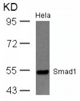
ARG51284 anti-Smad1 antibody WB image
Western Blot: extracts from HeLa cells stained with anti-Smad1 antibody ARG51284.
-
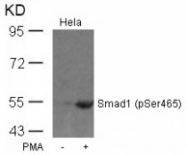
ARG51794 anti-Smad1 phospho (Ser465) antibody WB image
Western Blot: extracts from HeLa cells untreated or treated with PMA stained with anti-Smad1 (phospho Ser465) antibody ARG51794.
-
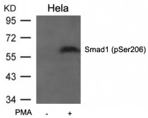
ARG51848 anti-Smad1 phospho (Ser206) antibody WB image
Western Blot: extracts from HeLa cells untreated or treated with PMA stained with anti-Smad1 (phospho Ser206) antibody ARG51848.
-
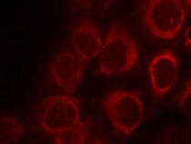
ARG51284 anti-Smad1 antibody ICC/IF image
Immunofluorescence: methanol-fixed HeLa cells stained with anti-Smad1 antibody ARG51284.
-
ARG65351 Goat anti-Rabbit IgG antibody (HRP) WB image
Western blot: Rat placental stained with ARG57589 anti-MTNR1A antibody at 1:1000 dilution, ARG65351 Goat anti-Rabbit IgG antibody (HRP) at 1:5000 dilution.
From Jinzhi Li et al. J Reprod Immunol. (2023), doi: 10.1016/j.jri.2023.104166, Fig. 2.B.
-
ARG65351 Goat anti-Rabbit IgG antibody (HRP) WB image
Western blot: Mouse retina stained with ARG65693 anti-alpha Tubulin antibody and ARG65351 Goat anti-Rabbit IgG antibody (HRP)
From Xiaoyuan Ye et al. Mol Ther Nucleic Acids. (2024), doi: 10.1016/j.omtn.2024.102209, Fig. 5.D.
-
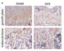
ARG65351 Goat anti-Rabbit IgG antibody (HRP) IHC-P image
From Yu-Qian Song et al. J Mol Med (Berl) (2022), doi: 10.1007/s00109-021-02165-0, Fig. 5.c.
