ARG30240
Phospho IGF1 Receptor Antibody Duo (Total, pY1161)
Cancer antibody; Developmental Biology antibody; Neuroscience antibody; Signaling Transduction antibody
Component
| Cat No | Component Name | Host clonality | Reactivity | Application | Package |
|---|---|---|---|---|---|
| ARG51590 | anti-IGF1 Receptor phospho (Tyr1161) antibody | Rabbit pAb | Hu, Ms, Rat | ICC/IF, IHC-P, WB | 50 μl |
| ARG51076 | anti-IGF1 Receptor antibody | Rabbit pAb | Hu, Ms, Rat | BL, FACS, ICC/IF, IHC-P, WB | 50 μl |
Overview
| Product Description | IGF-1R is a receptor tyrosine kinase which mediates actions of insulin-like growth factor 1 (IGF1). It binds IGF1 with higher affinity and IGF2 and insulin with a lower affinity. The activated IGF-1R is involved in cell growth and survival control. IGF1R is crucial for tumor transformation and survival of malignant cell. Ligand binding on IGF-1R activates the receptor kinase, leading to receptor autophosphorylation, and tyrosines phosphorylation of multiple substrates including the insulin-receptor substrates (IRS1/2), Shc and 14-3-3 proteins. Phosphorylation of IRS proteins lead to the activation of two main signaling pathways: the PI3K-AKT/PKB pathway and the Ras-MAPK pathway. The result of activating the MAPK pathway is increased cellular proliferation, whereas activating the PI3K pathway inhibits apoptosis and stimulates protein synthesis. Vazquez-Martin et al. 2013. Sci Rep 3: 2560 Lovly et al. 2014. Nat Med 20(9): 1027-34 Related news: Tumor microenvironments are shown to affect progression of several cancer subtypes |
|---|---|
| Target Name | IGF1 Receptor |
| Antigen Species | Human |
| Alternate Names | Phospho IGF1 Receptor antibody; Phospho Insulin-like growth factor 1 receptor antibody; Phospho IGF1R antibody; IGF1 Receptor antibody; IGF1 Receptor phospho (Tyr1161) antibody |
Properties
| Storage Instruction | For continuous use, store undiluted antibody at 2-8°C for up to a week. For long-term storage, aliquot and store at -20°C or below. Storage in frost free freezers is not recommended. Avoid repeated freeze/thaw cycles. Suggest spin the vial prior to opening. The antibody solution should be gently mixed before use. |
|---|---|
| Note | For laboratory research only, not for drug, diagnostic or other use. |
Bioinformation
| Gene Symbol | IGF1R |
|---|---|
| Gene Full Name | Phospho Insulin-like growth factor 1 receptor (IGF1R) Antibody Duo |
| Background | This receptor binds insulin-like growth factor with a high affinity. It has tyrosine kinase activity. The insulin-like growth factor I receptor plays a critical role in transformation events. Cleavage of the precursor generates alpha and beta subunits. It is highly overexpressed in most malignant tissues where it functions as an anti-apoptotic agent by enhancing cell survival. Alternatively spliced transcript variants encoding distinct isoforms have been found for this gene. [provided by RefSeq, May 2014] |
| Function | Receptor tyrosine kinase which mediates actions of insulin-like growth factor 1 (IGF1). Binds IGF1 with high affinity and IGF2 and insulin (INS) with a lower affinity. The activated IGF1R is involved in cell growth and survival control. IGF1R is crucial for tumor transformation and survival of malignant cell. Ligand binding activates the receptor kinase, leading to receptor autophosphorylation, and tyrosines phosphorylation of multiple substrates, that function as signaling adapter proteins including, the insulin-receptor substrates (IRS1/2), Shc and 14-3-3 proteins. Phosphorylation of IRSs proteins lead to the activation of two main signaling pathways: the PI3K-AKT/PKB pathway and the Ras-MAPK pathway. The result of activating the MAPK pathway is increased cellular proliferation, whereas activating the PI3K pathway inhibits apoptosis and stimulates protein synthesis. Phosphorylated IRS1 can activate the 85 kDa regulatory subunit of PI3K (PIK3R1), leading to activation of several downstream substrates, including protein AKT/PKB. AKT phosphorylation, in turn, enhances protein synthesis through mTOR activation and triggers the antiapoptotic effects of IGFIR through phosphorylation and inactivation of BAD. In parallel to PI3K-driven signaling, recruitment of Grb2/SOS by phosphorylated IRS1 or Shc leads to recruitment of Ras and activation of the ras-MAPK pathway. In addition to these two main signaling pathways IGF1R signals also through the Janus kinase/signal transducer and activator of transcription pathway (JAK/STAT). Phosphorylation of JAK proteins can lead to phosphorylation/activation of signal transducers and activators of transcription (STAT) proteins. In particular activation of STAT3, may be essential for the transforming activity of IGF1R. The JAK/STAT pathway activates gene transcription and may be responsible for the transforming activity. JNK kinases can also be activated by the IGF1R. IGF1 exerts inhibiting activities on JNK activation via phosphorylation and inhibition of MAP3K5/ASK1, which is able to directly associate with the IGF1R. When present in a hybrid receptor with INSR, binds IGF1. PubMed:12138094 shows that hybrid receptors composed of IGF1R and INSR isoform Long are activated with a high affinity by IGF1, with low affinity by IGF2 and not significantly activated by insulin, and that hybrid receptors composed of IGF1R and INSR isoform Short are activated by IGF1, IGF2 and insulin. In contrast, PubMed:16831875 shows that hybrid receptors composed of IGF1R and INSR isoform Long and hybrid receptors composed of IGF1R and INSR isoform Short have similar binding characteristics, both bind IGF1 and have a low affinity for insulin. [UniProt] |
| Highlight | Related Product: anti-IGF1 Receptor antibody; |
| Research Area | Cancer antibody; Developmental Biology antibody; Neuroscience antibody; Signaling Transduction antibody |
| PTM | Autophosphorylated on tyrosine residues in response to ligand binding. Autophosphorylation occurs in trans, i.e. one subunit of the dimeric receptor phosphorylates tyrosine residues on the other subunit. Autophosphorylation occurs in a sequential manner; Tyr-1165 is predominantly phosphorylated first, followed by phosphorylation of Tyr-1161 and Tyr-1166. While every single phosphorylation increases kinase activity, all three tyrosine residues in the kinase activation loop (Tyr-1165, Tyr-1161 and Tyr-1166) have to be phosphorylated for optimal activity. Can be autophosphorylated at additional tyrosine residues (in vitro). Autophosphorylated is followed by phosphorylation of juxtamembrane tyrosines and C-terminal serines. Phosphorylation of Tyr-980 is required for IRS1- and SHC1-binding. Phosphorylation of Ser-1278 by GSK-3beta restrains kinase activity and promotes cell surface expression, it requires a priming phosphorylation at Ser-1282. Dephosphorylated by PTPN1 (By similarity). Polyubiquitinated at Lys-1168 and Lys-1171 through both 'Lys-48' and 'Lys-29' linkages, promoting receptor endocytosis and subsequent degradation by the proteasome. Ubiquitination is facilitated by pre-existing phosphorylation. Sumoylated with SUMO1. Controlled by regulated intramembrane proteolysis (RIP). Undergoes metalloprotease-dependent constitutive ectodomain shedding to produce a membrane-anchored 52 kDa C-Terminal fragment which is further processed by presenilin gamma-secretase to yield an intracellular 50 kDa fragment. |
Images (7) Click the Picture to Zoom In
-

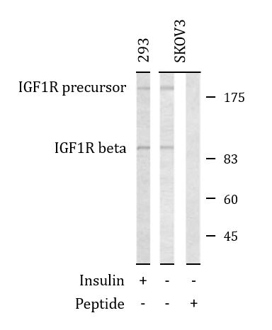
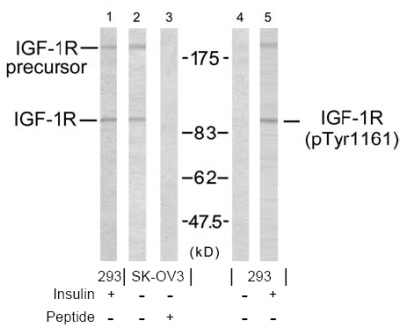
ARG51076 anti-IGF1 Receptor antibody WB image
Western blot: 293 and SKOV3 cells treated or un-treated with insulin and stained with ARG51076 anti-IGF1 Receptor antibody. Lane 3 also pre-incubated with the peptides used for antibody raising.
-

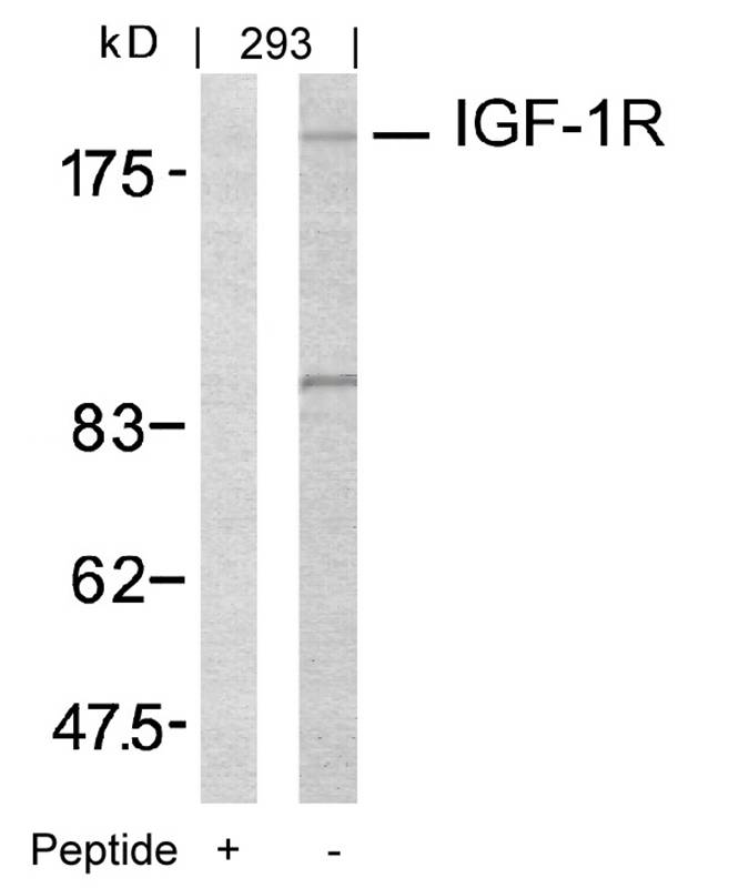
ARG51076 anti-IGF-1R antibody WB validated image
Western Blot: extracts from 293 cells stained with anti-IGF-1R antibody ARG51076 and the same antibody preincubated with blocking peptide.
-

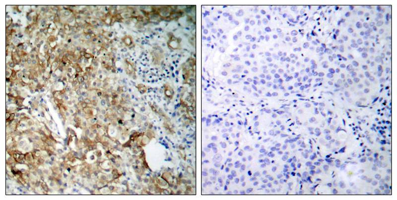
ARG51076 anti-IGF-1R antibody IHC-P validated image
Immunohistochemistry: paraffin-embedded human breast carcinoma tissue stained with anti-IGF-1R antibody ARG51076 (left) or the same antibody preincubated with blocking peptide (right).
-
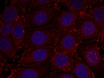
ARG51076 anti-IGF-1R antibody ICC/IF validated image
Immunofluorescence: methanol-fixed MCF cells stained with anti-IGF-1R antibody ARG51076.
-

ARG51590 anti-IGF-1R (phospho Tyr1161) antibody WB validated image
Western blot: 293 and SKOV3 cells treated or un-treated with insulin stained with IGF-1R antibody (Line 1, 2 and 3) and IGF-1R (phospho-Tyr1161) antibody (ARG51590, Line 4 and 5). Line3 and Line4 also pre-incubated with the peptides used for antibody raising.
-

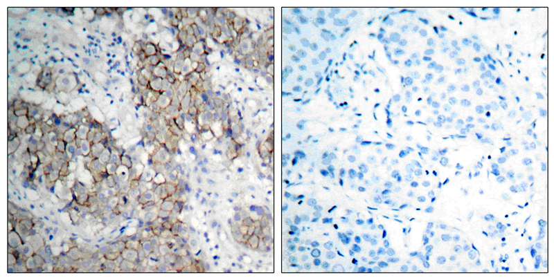
ARG51590 anti-IGF-1R (phospho Tyr1161) antibody IHC-P validated image
Immunohistochemistry: paraffin- embedded human breast carcinoma tissue stained with anti-IGF-1R (phospho Tyr1161) antibody ARG51590).
-
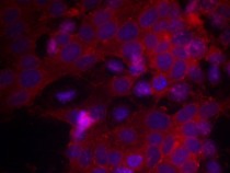
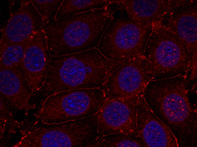
ARG51590 anti-IGF-1R (phospho Tyr1161) antibody ICC/IF validated image
Immunofluorescence: methanol-fixed MCF7 cells stained with anti-IGF-1R (phospho Tyr1161) antibody ARG51590, Red).

Specific References
Role of Extracellular Vimentin in Cancer-Cell Functionality and Its Influence on Cell Monolayer Permeability Changes Induced by SARS-CoV-2 Receptor Binding Domain.
ARG51076: Blocking / Human


电针对控制性超排卵小鼠子宫内膜磷酸化胰岛素样生长因子-1受体表达及其信号转导通路的调控
ARG51076, ARG51590: WB / Mouse


