ARG30238
Phospho EGFR Antibody Duo (Total, pY992)
Cancer antibody; Signaling Transduction antibody
Component
| Cat No | Component Name | Host clonality | Reactivity | Application | Package |
|---|---|---|---|---|---|
| ARG10518 | anti-EGFR antibody [EGFR1] | Mouse mAb | Hu, Ms, Hrs | FACS, ICC/IF, IHC-Fr, IP, WB | 50 μg |
| ARG53931 | anti-EGFR phospho (Tyr992) antibody [EM-12] | Mouse mAb | Hu | FACS, IP, WB | 50 μg |
Overview
| Target Name | EGFR |
|---|---|
| Alternate Names | Phospho EGFR antibody; Phospho Epidermal growth factor receptor antibody; EGFR antibody; EGFR phospho (Tyr992) antibody |
Properties
| Storage Instruction | For continuous use, store undiluted antibody at 2-8°C for up to a week. For long-term storage, aliquot and store at -20°C or below. Storage in frost free freezers is not recommended. Avoid repeated freeze/thaw cycles. Suggest spin the vial prior to opening. The antibody solution should be gently mixed before use. |
|---|---|
| Note | For laboratory research only, not for drug, diagnostic or other use. |
Bioinformation
| Gene Symbol | EGFR |
|---|---|
| Gene Full Name | Phospho Epidermal growth factor receptor (EGFR) Antibody Duo |
| Background | EGFR is a transmembrane glycoprotein that is a member of the protein kinase superfamily. This protein is a receptor for members of the epidermal growth factor family. EGFR is a cell surface protein that binds to epidermal growth factor. Binding of the protein to a ligand induces receptor dimerization and tyrosine autophosphorylation and leads to cell proliferation. Mutations in this gene are associated with lung cancer. Multiple alternatively spliced transcript variants that encode different protein isoforms have been found for this gene. [provided by RefSeq, Jul 2010] |
| Function | Receptor tyrosine kinase binding ligands of the EGF family and activating several signaling cascades to convert extracellular cues into appropriate cellular responses. Known ligands include EGF, TGFA/TGF-alpha, amphiregulin, epigen/EPGN, BTC/betacellulin, epiregulin/EREG and HBEGF/heparin-binding EGF. Ligand binding triggers receptor homo- and/or heterodimerization and autophosphorylation on key cytoplasmic residues. The phosphorylated receptor recruits adapter proteins like GRB2 which in turn activates complex downstream signaling cascades. Activates at least 4 major downstream signaling cascades including the RAS-RAF-MEK-ERK, PI3 kinase-AKT, PLCgamma-PKC and STATs modules. May also activate the NF-kappa-B signaling cascade. Also directly phosphorylates other proteins like RGS16, activating its GTPase activity and probably coupling the EGF receptor signaling to the G protein-coupled receptor signaling. Also phosphorylates MUC1 and increases its interaction with SRC and CTNNB1/beta-catenin. Isoform 2 may act as an antagonist of EGF action. [Uniprot] |
| Highlight | Related Product: anti-EGFR antibody; anti-EGFR phospho (Tyr992) antibody; |
| Research Area | Cancer antibody; Signaling Transduction antibody |
| PTM | Phosphorylation at Ser-695 is partial and occurs only if Thr-693 is phosphorylated. Phosphorylation at Thr-678 and Thr-693 by PRKD1 inhibits EGF-induced MAPK8/JNK1 activation. Dephosphorylation by PTPRJ prevents endocytosis and stabilizes the receptor at the plasma membrane. Autophosphorylation at Tyr-1197 is stimulated by methylation at Arg-1199 and enhances interaction with PTPN6. Autophosphorylation at Tyr-1092 and/or Tyr-1110 recruits STAT3. Dephosphorylated by PTPN1 and PTPN2. Monoubiquitinated and polyubiquitinated upon EGF stimulation; which does not affect tyrosine kinase activity or signaling capacity but may play a role in lysosomal targeting. Polyubiquitin linkage is mainly through 'Lys-63', but linkage through 'Lys-48', 'Lys-11' and 'Lys-29' also occurs. Deubiquitination by OTUD7B prevents degradation. Ubiquitinated by RNF115 and RNF126 (By similarity). Methylated. Methylation at Arg-1199 by PRMT5 stimulates phosphorylation at Tyr-1197. |
Images (3) Click the Picture to Zoom In
-
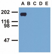
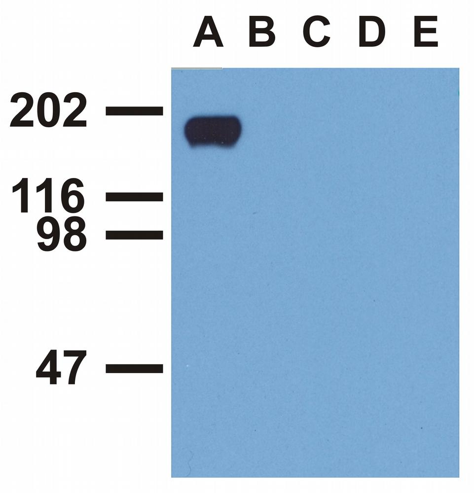
Monoclonal antibody clone EM-12 WB validated image
Western Blot: A. EGF-treated A431 cell lysate, B. CALU-3 cell lysate, C. MCF-7 cell lysate, D. Jurkat cell lysate, E. RAMOS cell lysate stained with antibody clone EM-12
-
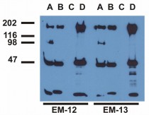
Monoclonal antibody clone EM-12 Immunoprecipitation validated image
Immunoprecipitation: EGF-treated A431 cell was immunoprecipitated with (A) EM-12 antibody, (B) EM-13 antibody, (C) anti-EGFR commercial polyclonal antibody, (D) anti-EGFR monoclonal mAb108 antibody. The precipitates were stained with EM-12 or EM-13 antibody.
-
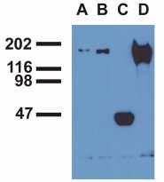
Monoclonal antibody clone EM-12 Immunoprecipitation validated image
Immunoprecipitation: EGF-treated A431 cell was immunoprecipitated with (A) EM-12 antibody, (B) EM-13 antibody, (C) anti-EGFR commercial polyclonal antibody, (D) anti-EGFR monoclonal mAb108 antibody. The precipitates were stained with commercial anti-EGFR polyclonal antibody.