ARG82522
Mouse SLIT2 ELISA Kit
Mouse SLIT2 ELISA Kit for ELISA and Mouse
Component
| Cat No | Component Name | Package | Temp |
|---|---|---|---|
| ARG82522-001 | Antibody-coated microplate | 8 X 12 strips | 4°C. Unused strips should be sealed tightly in the air-tight pouch. |
| ARG82522-002 | Standard | 2 X 10 ng/vial | 4°C |
| ARG82522-003 | Standard/Sample diluent | 30 ml (Ready to use) | 4°C |
| ARG82522-004 | Antibody conjugate concentrate (100X) | 1 vial (100 µl) | 4°C |
| ARG82522-005 | Antibody diluent buffer | 12 ml (Ready to use) | 4°C |
| ARG82522-006 | HRP-Streptavidin concentrate (100X) | 1 vial (100 µl) | 4°C |
| ARG82522-007 | HRP-Streptavidin diluent buffer | 12 ml (Ready to use) | 4°C |
| ARG82522-008 | 25X Wash buffer | 20 ml | 4°C |
| ARG82522-009 | TMB substrate | 10 ml (Ready to use) | 4°C (Protect from light) |
| ARG82522-010 | STOP solution | 10 ml (Ready to use) | 4°C |
| ARG82522-011 | Plate sealer | 4 strips | Room temperature |
Overview
| Product Description | ARG82522 Mouse SLIT2 ELISA Kit is an Enzyme Immunoassay kit for the quantification of Mouse SLIT2 in serum, plasma (EDTA, heparin) and cell culture supernatants. |
|---|---|
| Tested Reactivity | Ms |
| Tested Application | ELISA |
| Target Name | SLIT2 |
| Conjugation | HRP |
| Conjugation Note | Substrate: TMB and read at 450 nm. |
| Sensitivity | 78 pg/ml |
| Sample Type | Serum, plasma (EDTA, heparin) and cell culture supernatants. |
| Standard Range | 156 - 10000 pg/ml |
| Sample Volume | 100 µl |
| Precision | Intra-Assay CV: 5.3% Inter-Assay CV: 6.9% |
| Alternate Names | Slit-2; Slit homolog 2 protein; SLIL3 |
Application Instructions
| Assay Time | ~ 5 hours |
|---|
Properties
| Form | 96 well |
|---|---|
| Storage Instruction | Store the kit at 2-8°C. Keep microplate wells sealed in a dry bag with desiccants. Do not expose test reagents to heat, sun or strong light during storage and usage. Please refer to the product user manual for detail temperatures of the components. |
| Note | For laboratory research only, not for drug, diagnostic or other use. |
Bioinformation
| Database Links | |
|---|---|
| Gene Symbol | SLIT2 |
| Gene Full Name | slit guidance ligand 2 |
| Background | This gene encodes a member of the slit family of secreted glycoproteins, which are ligands for the Robo family of immunoglobulin receptors. Slit proteins play highly conserved roles in axon guidance and neuronal migration and may also have functions during other cell migration processes including leukocyte migration. Members of the slit family are characterized by an N-terminal signal peptide, four leucine-rich repeats, nine epidermal growth factor repeats, and a C-terminal cysteine knot. Proteolytic processing of this protein gives rise to an N-terminal fragment that contains the four leucine-rich repeats and five epidermal growth factor repeats and a C-terminal fragment that contains four epidermal growth factor repeats and the cysteine knot. Both full length and cleaved proteins are secreted extracellularly and can function in axon repulsion as well as other specific processes. Alternative splicing results in multiple transcript variants. [provided by RefSeq, Sep 2015] |
| Function | Thought to act as molecular guidance cue in cellular migration, and function appears to be mediated by interaction with roundabout homolog receptors. During neural development involved in axonal navigation at the ventral midline of the neural tube and projection of axons to different regions. SLIT1 and SLIT2 seem to be essential for midline guidance in the forebrain by acting as repulsive signal preventing inappropriate midline crossing by axons projecting from the olfactory bulb. In spinal chord development may play a role in guiding commissural axons once they reached the floor plate by modulating the response to netrin. In vitro, silences the attractive effect of NTN1 but not its growth-stimulatory effect and silencing requires the formation of a ROBO1-DCC complex. May be implicated in spinal chord midline post-crossing axon repulsion. In vitro, only commissural axons that crossed the midline responded to SLIT2. In the developing visual system appears to function as repellent for retinal ganglion axons by providing a repulsion that directs these axons along their appropriate paths prior to, and after passage through, the optic chiasm. In vitro, collapses and repels retinal ganglion cell growth cones. Seems to play a role in branching and arborization of CNS sensory axons, and in neuronal cell migration. In vitro, Slit homolog 2 protein N-product, but not Slit homolog 2 protein C-product, repels olfactory bulb (OB) but not dorsal root ganglia (DRG) axons, induces OB growth cones collapse and induces branching of DRG axons. Seems to be involved in regulating leukocyte migration. [UniProt] |
| Cellular Localization | Secreted. Note=The C-terminal cleavage protein is more diffusible than the larger N-terminal protein that is more tightly cell associated. [UniProt] |
| Highlight | Related products: SLIT2 antibodies; SLIT2 ELISA Kits; New ELISA data calculation tool: Simplify the ELISA analysis by GainData |
Images (1) Click the Picture to Zoom In
-

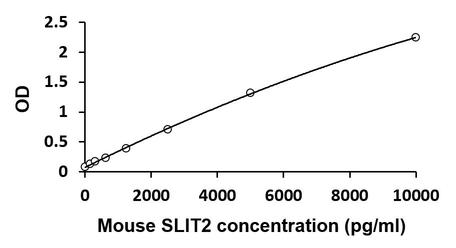
ARG82522 Mouse SLIT2 ELISA Kit standard curve image
ARG82522 Mouse SLIT2 ELISA Kit results of a typical standard run with optical density reading at 450 nm.
| Title | Download Link |
|---|---|
| ARG82522 Mouse SLIT2 ELISA Kit User manual |
 Download Download
|


