ARG30162
Cytotoxic T Cell Surface Marker Antibody Panel (FACS)
Cell Biology and Cellular Response antibody; Developmental Biology antibody; Immune System antibody; Neuroscience antibody; Signaling Transduction antibody
Component
| Cat No | Component Name | Host clonality | Reactivity | Application | Package |
|---|---|---|---|---|---|
| ARG62928 | anti-CD8 antibody [MEM-31] | Mouse mAb | Hu | CyTOF®-candidate, FACS, IP | 50 μg |
| ARG62889 | anti-CD54 / ICAM1 antibody [1H4] (FITC) | Mouse mAb | Hu | FACS | 50 tests |
| ARG53814 | anti-CD28 antibody [CD28.2] (APC) | Mouse mAb | Hu, NHuPrm | FACS | 50 tests |
| ARG62855 | anti-CD45 antibody [MEM-28] (Biotin) | Mouse mAb | Hu | FACS | 50 μg |
Overview
| Product Description | A cytotoxic T cell is a T lymphocyte that kills cancer cells, virally infected cells and cells that are under damage. Most T lymphocytes express a subset of surface markers such as CD8, CD45 and CD54. CD28 expresses on the surface of T cells and provide co-stimulatory signals required for T cell activation. |
|---|---|
| Target Name | Cytotoxic T Cell Surface Marker |
| Alternate Names | Cytotoxic T Cell Surface Marker antibody; APC-conjugated CD28 antibody; Biotin-conjugated CD45 antibody; FITC-conjugated CD54 / ICAM1 antibody; CD8 antibody |
Properties
| Storage Instruction | Store antibodies at 4°C or -20°C. Please refer to the each product datasheet for detail temperatures of the antibodies. |
|---|---|
| Note | For laboratory research only, not for drug, diagnostic or other use. |
Bioinformation
| Gene Full Name | Antibody Panel for Cytotoxic T Cell Surface Marker |
|---|---|
| Highlight | Related Product: anti-CD8 antibody; anti-CD54 / ICAM1 antibody; anti-CD28 antibody; anti-CD45 antibody; |
| Research Area | Cell Biology and Cellular Response antibody; Developmental Biology antibody; Immune System antibody; Neuroscience antibody; Signaling Transduction antibody |
Images (9) Click the Picture to Zoom In
-
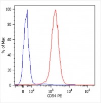
Monoclonal antibody clone 1H4 Flow Cytometry analysis image
Flow Cytometry: U937 human histiocytic lymphoma cell stained with antibody clone 1H4.
Total viable cells were used for analysis. -
Monoclonal antibody clone CD28.2 Flow Cytometry analysis image
Flow Cytometry: Human peripheral blood leukocytes stained with antibody clone CD28.2.
-
Monoclonal antibody clone MEM-28 Flow Cytometry analysis image
Flow Cytometry: Human peripheral blood cells stained with antibody clone MEM-28.
-
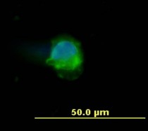
Monoclonal antibody clone MEM-28 ICC/IF image
Immunofluorescence: Human peripheral blood mononuclear cell stained with clone MEM-28 (green)
Cell nuclei was stained with DAPI (blue). -
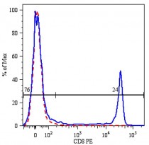
Monoclonal antibody clone MEM-31 Flow Cytometry analysis image
Flow Cytometry: Human peripheral blood cells stained with antibody clone MEM-31.
-
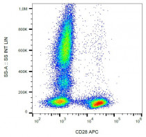
ARG53814 anti-CD28 antibody [CD28.2] (APC) FACS image
Flow Cytometry: Human peripheral blood leukocytes stained with ARG53814 anti-CD28 antibody [CD28.2] (APC).
-
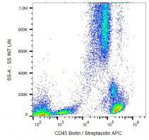
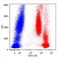
ARG62855 anti-CD45 antibody [MEM-28] (Biotin) FACS image
Flow Cytometry: Human peripheral blood cells stained with ARG62855 anti-CD45 antibody [MEM-28] (Biotin), followed by Streptavidin (APC).
-
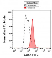
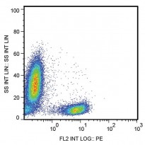
ARG62889 anti-CD54 / ICAM1 antibody [1H4] (FITC) FACS image
Flow Cytometry: Separation of Human CD54 positive Monocytes (red) from Human CD54 negative Lymphocytes (black-dashed). Human peripheral blood stained with ARG62889 anti-CD54 / ICAM1 antibody [1H4] (FITC).
-
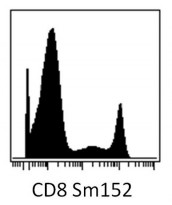
ARG62928 anti-CD8 antibody [MEM-31] CyTOF image
CyTOF: PBMC (after Ficoll-Paque separation) stained with ARG62928 anti-CD8 antibody [MEM-31] (Sm152). Singlet cells were gated for data analysis.


