Microglial help TAM-ing inflammation in the brain

Microglial help TAM-ing inflammation in the brain


Some cells are meant to live, and some are meant to die. Tens of thousands of cells are undergoing apoptosis in our body each day without being noticed or identified. This is because the dead cells are quickly cleared up by the TAM receptor-mediated phagocytosis the moment apoptotic cells started to display Phosphatidylserine (PtdSer) on the outer cell membrane.

The TAM receptors –Tyro3, AXL, and Mer- comprise a unique family of receptor tyrosine kinases, who play no essential role in embryonic development, but function as homeostasis regulators in adult tissues that are continually subjected to challenge and renewal throughout life. TAM ligands (Gas6 or Pros1) serves as a bridging molecule that links a TAM receptor, expressed on the surface of phagocyte, to PtdSer, which is displayed on the surface of apoptotic cells to be engulfed and broken down. Deficiencies in TAM signaling are thought to contribute to chronic inflammation and autoimmune diseases, and overexpression of TAMs has been shown to affect cancer progression and metastasis.


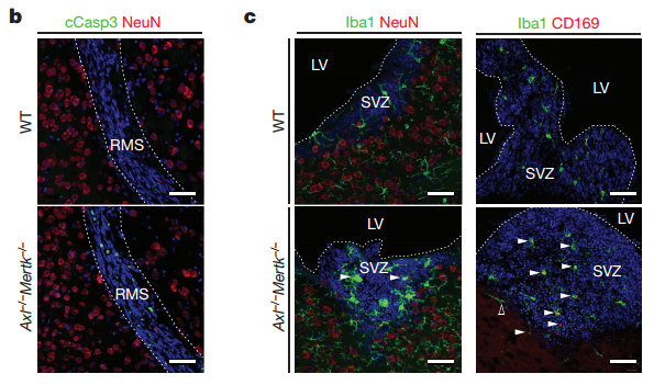
Now, in a research published in Nature, scientists found that TAM receptor also helps to regulate microglial functions as they patrol through the brain and spinal cord to identify apoptotic cells in order to maintain homeostasis in the central nervous system. G. Lemke and colleagues reported that adult mice lacking microglial Mer and AXL exhibit marked accumulation of apoptotic cells in the neurogenic region of CNS. Besides, microglial expression of AXL was upregulated in mouse model of Parkinson’s disease, and the loss of Mer and AXL in these mice modestly prolonged and extended their survival.


(From original article Figure 1, Fourgeaud et al. (2016) Nature)


The research of TAM signaling in neurodegenerative disease such as Parkinson’s offers new insight into identifying potential targets for diagnostic and therapeutic intervention in these diseases. arigo provides quality antibodies and antibody duos to help you accelerate and excel in your research.


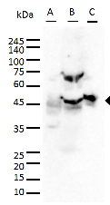
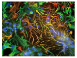
GFAP antibody (ARG10122, ARG52312)
ARG10122  

ARG52312


A: U87MG
B: Mouse brain
C: Rat brain
  Cultured neurons and glia


Iba antibody (ARG63338)
 

Mouse brain


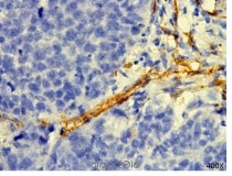
CD31 (ARG52748)
     
 
MDA-MB-231
(tumor xenograft study)
 

HUVEC cells


ARG52224
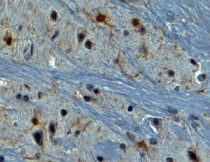
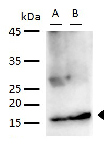
Alpha Synuclein
 

ARG51667
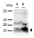
Alpha Synuclein pS129


A:Mouse brain
B: Rat brain
  A:Mouse brain
B: Rat brain