Research solutions for Macrophages
Research solutions for Macrophages
Macrophages are specialized cells involved in defense by engulfing pathogens, microbes, harmful cells, and cellular debris.

When sensing pathogens, toxins or DAMPs (damage-associated molecular patterns), macrophages trigger inflammation by releasing inflammatory cytokines that activate more immune cells. Besides, macrophages also present antigens to T cells, thus helping to initiate adaptive immunity. Together with neutrophils, macrophages are the cellular hallmarks of chronic inflammation. In addition, macrophages also participate in tissue repair.
Macrophages are classified into M1 and M2 subtypes that mediate inflammation and repair respectively. Circulating monocytes are recruited to tissues, where they differentiate to naïve macrophages (M0) and then polarize toward inflammatory M1 or resolving M2 phenotypes in response to different stimuli.
|
|
|
|
|
|
|
Mouse M1 / M2 Cytokines Multiplex ELISA Kit (ARG82913) ● M1 cytokines: IL-6, TNF-α ● M2 cytokines: IL-4, IL-10 |
|
M1 / M2 / TAM Marker Antibody Panel (ARG30333) 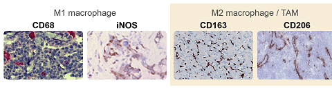 |
Tumor-associated macrophages (TAMs), primarily of the M2 type, are the most abundant immune cell population in tumors, providing an immunosuppressive niche that supports tumor progression. TAMs inhibit the cytotoxic activity of cytotoxic T lymphocytes (CTLs) and natural killer (NK) cells by producing inhibitory factors IL-10, TGF-β, kynurenine, PGE2 …etc. These inhibitory factors also promote the expansion of immunosuppressive cells including regulatory T cells (Tregs) and myeloid-derived suppressor cells (MDSCs). In addition, TAMs induce vascularization of tumor tissue by producing VEGF, PDGF, and TGF-β. Depletion or reprogramming of TAMs toward an M1-type has shown potential for cancer therapy.
Inflammasomes are multiprotein complexes that regulate the maturation of secretion of pro-inflammatory cytokines IL-1β and IL-18. Monocytes and macrophages are the main cells expressing the inflammasome genes. Multiple reports reveal that NLRP3 inflammasome mediates M1 polarization. The association of NLRC4 inflammasome and M2 polarization has been reported. However, the mechanism of inflammasome-regulated macrophage polarization remain to be discovered.
