A non-autophatic role of Atg9a in necrosis and developmental bone formation
A non-autophatic role of Atg9a in necrosis and developmental bone formation
|
Atg9a is well known for its function on autophagy. Recently, a new role of Atg9a in necrosis and developmental bone formation has been reported. An article published on Nat. Commun. by Dr. Imagawa and his colleagues shows that the necrotic cell death occurred at the prospective bone surface in WT embryo while the necrotic cell death was disappeared from the prospective bone surface in Atg9a KO embryo. However, this necrotic death was found in Atg5 KO embryo, suggesting the autophagy does not have a role in the necrotic cell death at the prospective bone surface. Their finding suggests that Atg9a-dependent necrosis has an active role in bone surface formation during development. Let arigo’s excellent antibodies and reagents accelerate your research on necrosis and autophagy.
|
||
 |
 |
 |
| Necrosis | ||
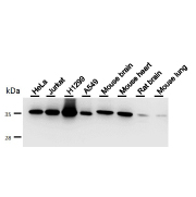
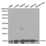
| Cyclophilin A antibody (ARG55463) | ||
 |
 |
 |
| Necroptosis | ||
.jpg) |
.jpg) |
.jpg) |
Reference:
Imagawa, Y. et al. (2016) Vital staining for cell death identifies Atg9a-dependent necrosis in developmental bone formation in mouse. Nat. Commun. 7:13391.
