Microglial help TAM-ing inflammation in the brain
Microglial help TAM-ing inflammation in the brain
|
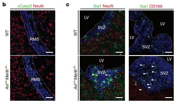
The TAM receptors –Tyro3, AXL, and Mer- comprise a unique family of receptor tyrosine kinases, who play no essential role in embryonic development, but function as homeostasis regulators in adult tissues that are continually subjected to challenge and renewal throughout life. TAM ligands (Gas6 or Pros1) serves as a bridging molecule that links a TAM receptor, expressed on the surface of phagocyte, to PtdSer, which is displayed on the surface of apoptotic cells to be engulfed and broken down. Deficiencies in TAM signaling are thought to contribute to chronic inflammation and autoimmune diseases, and overexpression of TAMs has been shown to affect cancer progression and metastasis.
|

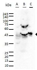
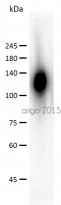
| GFAP antibody (ARG10122, ARG52312) | ||
| ARG10122 | ||
|
|
|
|
| A: U87MG B: Mouse brain C: Rat brain |
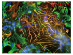
Cultured neurons and glia | |
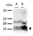
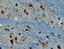
| Iba antibody (ARG63338) | ||
|
|
||
| Mouse brain | ||
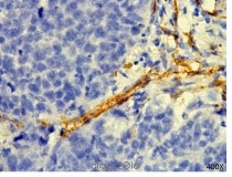
| CD31 (ARG52748) | ||
 |
 |
|
| MDA-MB-231 (tumor xenograft study) |
HUVEC cells |
|